The new method, discovered by scientists at the Max Planck Institute of Neurobiology in Martinsried, can help researchers recognize specific neural cells directly responsible for certain actions and behaviors in small fish. Like never before, the artificial stimulation of neurons can elicit predictable and desired responses. Comprehending the integral parts of brain circuitry is an essential factor in understanding the complexity of even the most basic neural functions.
Recent advances, specifically in microscopy and imaging, have progressed researchers’ ability to track brain activity better. Unfortunately, previous methods have not allowed observers to differentiate between neural activity caused by stimuli and the activity caused by the animals’ response to said stimuli. Utilizing optogenetics, scientists can now follow the route of neural commands that lead directly to actions without confusing it with other neuron activity, usually quite challenging due to the entirely through connectedness of neuron networks where an individual cell can cause waves of activity throughout the entire nervous system. Thanks to a new study by Herwig Baier’s team at the Max Planck Institute, two major obstacles have been removed. Finally, scientists can narrow in on cause and consequence to particular cells and also track that activity through the brain as it incites behavior.

Just years ago, Baier’s lab team was able to locate the part of a zebrafish brain that caused its tail to move. However, they had no ability to record the patterns of neural activity. Since then Institute scientists have created a system that allows 3-D photostimulation of desired neurons while simultaneously imaging the brain activity of zebrafish larva. “The zebrafish with it’s small, the translucent brain is ideal for our new method,” says the paper’s co-author Marco Dal Maschio.
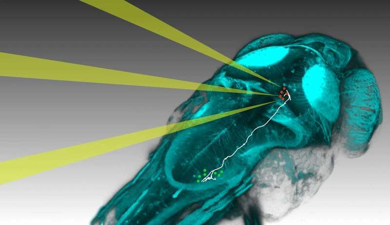
The first step to tracking activity was genetically engineering a zebrafish with photo-sensitive ion channels embedded in its nerve cells. Then, scientists could activate and control the fish with a light shined through their skulls. The team put these specialty larvae under a microscope and projected computer created images, holograms made of 3D light, into its brain. The infrared light beams were invisible to the larvae and arranged to activate a few unique neurons. The ensuing tail movement was captured by a speedy camera while the brain cells were stimulated with changing images. The experiment continued until researchers were able to identify the three neurons responsible for the bending tail. Then these cells were activated at the same time that quick 3D imaging recorded the activity throughout the neural network.
Using the dataset produced, a computer distinguished the associated patterns with different aspects of the resulting action (the fish bending its tail) and gave every responsible neuron a “contribution score”. At last, individual neurons with notable capacity were reproduced by creating its shape beneath a microscope. As their brains are quite similar to individual fish, a diagram of the circuitry can be drawn using the results from a number of comparable experiments.
“It’s the first time that a behavioral command can be traced as it spreads from a few initial cells throughout the brain to the actual physical action,” says Marco Dal Maschio optimistically, with good cause. Because of his and his lab mates new method, they can explore networks of neurons like never before. As brain structures have remained similar amongst fish and mammals, this new method and it’s resulting data will likely illuminate previously obscured neural activity and its relationship to physical behaviors in multiple species and not fish alone.
More News to Read


